


Peer Reviewed
Abstract
Photodisinfection therapy (PDT) is a treatment modality that involves the administration of a light-sensitive compound, known as a photosensitizer (PS), followed by light irradiation at a specific wavelength that excites or “activates” the PS. PDT is minimally invasive and already used clinically to treat a wide range of medical conditions. In its antimicrobial form, (antimicrobial PDT – aPDT) it has been shown to eradicate pathogenic microorganisms such as Gram-positive and Gram-negative bacteria, viruses, protozoa, and fungi and, unlike traditional antibiotics, does not induce resistance following repeated exposures to the therapy (Pedigo et al, 2009; Tavares et al, 2010; Costa et al, 2011; Cabiscol et al, 2000; Lauro et al, 2002; Jori & Coppellotti, 2007; Cassidy et al, 2010; Giuliani et al, 2010; Martins et al, 2018; Al-Mutairi et al, 2018).
For these reasons, we believe aPDT will evolve into an essential tool for infection control and become a vital part of the solution to the global AMR crisis. This report will underscore explain the fundamental principles of aPDT and illustrate the ways in which aPDT can be used to reduce the risk of hospital-acquired infections and improve patient outcomes.
Main Article
Fundamentals of Antimicrobial Photodisinfection Therapy (aPDT)
The first accounts of using light for the treatment of physical illness appeared in Egyptian, Indian, and Chinese writing more than 30 centuries ago (Deniell & Jill, 1991). The first detailed evidence for the antimicrobial activity of certain photosensitizers combined with light was documented in Munich (Raab, 1900), who noticed that the toxic effect of acridine dye on paramecia was greater on sunny days. Overshadowed by the development of antibiotics, another 80 years would pass before seminal work in aPDT began to appear in the literature (Bertoloni et al, 1985; Malik et al, 1990). The discovery and subsequent development of hematoporphyrin in 1841 is considered the most important event in the progress of aPDT (Diels & Arissian, 2011). Second and third-generation porphyrins, porphyrin derivatives, and benzoporphyrins have more recently been developed that improve the efficacy of aPDT (Abdel-Kader, 2016).
The basic electrodynamics (Figure 1) involved in photosensitized reactions involves the absorption of photons by the ground-state PS, causing electrons to be “pumped” to an excited state. This “activated” PS can then engage in many different kinds of chemical reactions that are destructive to microbes, such as electron transfer reactions and the formation of radicals, including the potent hydroxyl radical (Type I, redox reactions). A second activation pathway (Type II, peroxidation reactions) also exists, by which energy transfers in a resonant process from a long-lived PS triplet state to surrounding molecular oxygen, itself a ground-state triplet. The oxygen molecules in turn are pumped to their excited state, generating Reactive Oxygen Species (ROS): highly reactive chemical species including singlet oxygen, a powerful oxidizer capable of directly destroying microbes through lethal peroxidative reactions. It has been demonstrated that singlet oxygen can exert potent cytotoxic effects on microbes without being internalized (Dahl et al, 1987). The singlet oxygen lifetime in biological media is short – less than 0.05 µs – due to quenching by water, and therefore the mean diffusion distance of the molecule is less than 0.02 µm before returning to ground state (Moan & Berg, 1991). This short active lifetime localizes the kill to the immediate vicinity of the activated molecule.
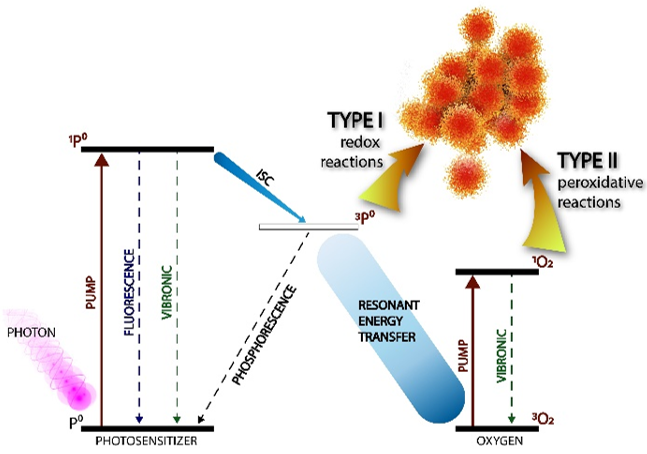
Figure 1: The electrodynamics of photodisinfection therapy. Source: Ondine Biomedical, Inc.
Depending on the chemical nature of the PS, its concentration, local fluid dynamic environment, pre-incubation time, and illumination time, the PS also localizes at different cellular targets. Studies with porphyrin photosensitizers have shown that at short pre-incubation and illumination times, the effects are limited to the microbial cell wall and cytoplasmic membrane, causing damage to transporter systems and transmembrane proteins, leading to cytoplasmic leakage (Malik et al, 1990). At moderate exposure times, a non-porphyrin PS can diffuse into the periplasmic space and damage the cytoplasmic membrane of Gram- negative microbes. Finally, at long exposure times, the PS can intercalate into (and damage) microbial DNA in the cytoplasmic compartment, both at the bacterial chromosome level and in the extrachromosomal plasmids (Valduga et al, 1993).
Photosensitizers are often positively charged to preferentially bind to negatively-charged microbial cell membranes. In contrast, human cells have both positive and negatively charged regions, but overall are electrically neutral. They take up less PS and therefore are more protected from damage (Loebel et al, 2016). The destructive reactions caused by singlet oxygen are therefore selective for the organisms to which the PS adheres. The destructive effect is further amplified by the PDT “bystander” effect (Alexandre et al, 2007) a cooperative inactivation process between cells in a given microcolony, most likely mediated by microbicidal photoproducts or the transfer of lysosomal enzymes from nearby cells.
aPDT for Treatment of Microbial Infection and Disease
The treatment of oral infections by aPDT has been extensively studied for many years and has become a well-established therapeutic option. Several recent reviews have demonstrated its efficacy for the treatment of periodontitis (Joseph et al, 2017; Azaripour et al, 2018; Meimandi et al, 2017), caries (Cieplik et al, 2017), endodontic infections (Mohammadi et al, 2017), and peri-implantitis (Ghanem et al, 2016). aPDT has also been found to be effective in the treatment of a variety of other infectious diseases caused by bacteria, fungi and protozoa including brain abscesses (Lombard et al, 1985), acne (Hongcharu et al, 2000; Wiegell et al, 2006; Tuchin et al, 2003; Seo et al, 2016; Tao et al, 2015; Serini et al, 2018), folliculitis (Lee et al, 2010), H. pylori (Wilder-Smith et al, 2002), diabetic and skin ulcers (Carrinho et al, 2018; Aspiroz et al, 2017; Lei et al, 2015; Morley et al, 2013; Mannucci et al, 2014), interdigital mycosis (Calzavara-Pinton et al, 2004), keratitis (Amescua et al, 2017), onychomycosis (Morgado et al, 2017), candidiasis (Scwingel et al, 2012), cutaneous leishmaniasis (Asilian & Davami, 2006), oral paracoccidiodomycosis (Dos Santos et al, 2017), and refractory chronic rhinosinusitis (Desrosiers et al, 2013). Treatment of viral infections with PDT also has a long clinical history. In the 1970s, a series of clinical studies demonstrated efficacy in treating infections due to the herpes simplex virus (Wainwright, 2003; Kharkwal et al, 2011). The most widely investigated viral infections have been those associated with human papilloma virus (HPV), a group of more than 150 types of virus that affect the skin and mucous membranes. In addition to causing diseases such as respiratory papillomatosis, genital warts, and skin warts (Ohtsuki et al, 2009; Hu et al, 2018), certain HPV types are carcinogenic and can result in cervical, vulvar, penile, and anal intraepithelial neoplasia (Grce & Mravak-Stipetić, 2014; Tommasino, 2014). aPDT with a variety of photosensitizers has been shown to be successful in the treatment of a range of HPV-associated infections including respiratory papillomatosis (Shikowitz et al, 1998; Shikowitz et al, 2005), plantar warts (Schroeter et al, 2005), condylomata acuminate (Wang et al, 2007; Shi et al, 2013), cervical intraepithelial neoplasia (Soergel et al, 2010; Hillemanns et al, 2014), and penile intraepithelial neoplasia (Paoli et al, 2006).
aPDT Is More Than a Microbicide
The damage inflicted by pathogenic microbes on their host, as well as their ability to avoid host defense systems, is mediated by a variety of virulence factors such as exotoxins, endotoxins, capsules, adhesins, invasins, and proteases (Casadevall & Pirofski, 2001). While antibiotics can kill microbes and thereby prevent further production of host-damaging molecules, extremely few have any effect on pre- existing virulence factors, which means that these molecules may have damaging effects even when the offending microbes have been killed (Lepper et al, 2002).
In contrast to most antibiotics, light-activated PSs are generally able to neutralize microbial virulence factors or reduce their effectiveness or decrease their expression. The ability to modify the biological activities of lipopolysaccharides (LPSs; i.e. endotoxin) is of particular interest because LPSs are potent immunomodulators that can induce secretion of several pro-inflammatory cytokines by host cells (Packer & Wilson, 2000; Pourhajibagher et al, 2018; Pourhajibagher et al, 2017; Shrestha et al, 2015; Giannelli et al, 2017; Tubby et al, 2009; Tseng et al, 2015; Bartolomeu et al, 2016; Calvino-Fernández et al, 2013; Pourhajibagher et al, 2016; Kato et al, 2013; Pereira et al, 2015; Cavaillon, 2018). Activated photosensitizers have been shown to be effective at reducing the activity of LPSs, proteases, and a variety of exotoxins. The ability of aPDT to not only kill the microbes responsible for an infection but also to inactivate or decrease the expression of many of the molecules responsible for host tissue destruction constitutes an important advantage over antibiotics as this combines both antimicrobial and anti- inflammatory approaches into a single treatment.
aPDT is Safe for Human Use
Numerous pre-clinical and clinical studies have demonstrated that aPDT is safe for use in treating infections in human tissues. For all the energetic reactivity of the ROS, several factors including extremely small time and distance scales, selectivity for anionic microbes, and the inherent resistance to oxidative stress of mammalian cells result in minimal damage to neighboring host tissues (Moan & Berg, 1991; Soukos et al, 1996; Millson et al, 1997; Soergel et al, 2010; Wang et al, 2007). Soukos et al. (1996) found that the viability of oral fibroblasts and keratinocytes was unaffected by the low concentration of Toluidine Blue O (TBO) and light dose needed to kill Streptococcus sanguinis. A number of photosensitizers, including MB and TBO, have been shown to have no deleterious effects on the gastric mucosa of rats at concentrations and light doses able to kill bacteria (Millson et al, 1997). In a clinical study aimed at detecting tissue damage associated with aPDT, two cycles of aPDT employing aminolevulinic acid esters as the PS were found to exert no damage to the cervix of the test patients (Soergel et al, 2010). The absence of tissue damage following the successful treatment of urethral condylomata acuminata (due to HPV) by aPDT using aminolevulinic acid has also been reported (Wang et al, 2007).
aPDT Does Not Induce Microbial Resistance
The generation of ROS in human immune cells (neutrophils, monocytes, and eosinophils) is one of the primary means by which our own immune system combats infecting microbes. It should therefore come as no surprise that highly-adaptable microbes have evolved protection strategies against these potent molecules by up-regulating antioxidant enzymes when exposed to ROS (Cabiscol et al, 2000), suggesting one method by which microbes could develop increased resistance to aPDT. However, numerous studies involving repeated exposure of microbes to aPDT and then re-testing the susceptibility of survivors have provided no evidence that resistance development occurs (Pedigo et al, 2009; Tavares et al, 2010; Costa et al, 2011; Cabiscol et al, 2000; Lauro et al, 2002; Jori & Coppellotti, 2007; Cassidy et al, 2010; Giuliani et al, 2010; Martins et al, 2018; Al-Mutairi et al, 2018). In particular, the speed of kill and the external mechanism of ROS appear to limit the ability to develop resistance to aPDT (Maisch, 2015).
In one example utilizing the PS Methylene Blue against MRSA, re-culturing experiments carried out over several consecutive years demonstrated no decrease in susceptibility to aPDT (Figure 2), whereas high-level resistance to oxacillin was established after less than a dozen cycles (Pedigo et al, 2009). This finding has been duplicated in studies with more complex sensitizers (Tavares et al, 2010) and also in viruses, where no increase in resistance was demonstrated after numerous cycles of aPDT (Costa et al, 2011).
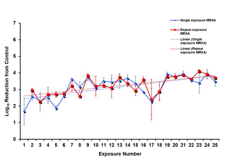
Figure 2: Repeated applications of aPDT to MRSA utilizing Methylene Blue does not promote microbial resistance. Source: Ondine Biomedical, Inc.
Conclusion
Photodisinfection Therapy is a well-known electrodynamic phenomenon that has become an established therapeutic option for a wide range of medical conditions – from microbial infections to cancer. More than 50 years of clinical use in humans has firmly established the fundamental safety and efficacy of myriad applications of this technology. In particular, antimicrobial PDT has tremendous potential to help combat the current global antimicrobial resistance crisis due to its demonstrated clinical efficacy and lack of resistance formation, all while promoting stewardship of existing antibiotics that are increasingly challenging and expensive to develop. aPDT is indeed an essential technology for both the present and the future of infection control.
References
Abdel-Kader, M. H. (2016). Antimicrobial Photodynamic Therapy: A Decade Of Development and Clinical Study. In Photodynamic Medicine, From Bench to Clinic. Ed. Kostron, H. and Hasan T., Royal Society of Chemistry, 1-21.
Alexandre, J., Hu, Y., Lu, W., Pelicano, H., & Huang, P. (2007). Novel action of paclitaxel against cancer cells: bystander effect mediated by reactive oxygen species. Cancer Research, 67:3512–3517.
Allison, R. R., Sibata, C., & Gay, H. (2009). PDT for cancers of the head and neck. Photodiagnosis and Photodynamic Therapy, 6(1), 1-2.
Al-Mutairi, R., Tovmasyan, A., Batinic-Haberle, I., & Benov, L. (2018). Sublethal photodynamic treatment does not lead to development of resistance. Frontiers in Microbiology, Jul 31;9:1699.
Amescua. G., Arboleda, A., Nikpoor, N., Durkee, H., Relhan, N., Aguilar, M. C., Flynn, H. W., Miller, D., Parel, J. M. (2017). Rose bengal photodynamic antimicrobial therapy: A novel treatment for resistant fusarium keratitis. Cornea, Sep;36(9):1141-1144.
Asilian, A., & Davami, M. (2006). Comparison between the efficacy of photodynamic therapy and topical paromomycin in the treatment of Old World cutaneous leishmaniasis: a placebo-controlled, randomized clinical trial. Clinical and Experimental Dermatology, Sep,31(5):634-7.
Aspiroz, C., Sevil, M., Toyas, C., & Gilaberte, Y. (2017). Photodynamic Therapy With Methylene Blue for Skin Ulcers Infected With Pseudomonas aeruginosa and Fusarium spp. Actas Dermo-Sifiliográficas, Jul-Aug;108(6):e45-e48.
Azaripour, A., Dittrich, S., Van Noorden, C. J. F., Willershausen, B. (2018). Efficacy of photodynamic therapy as adjunct treatment of chronic periodontitis: a systematic review and meta-analysis. Lasers in Medical Science, Feb;33(2):407-423.
Bader, M. J., Stepp, H., Beyer, W., Pongratz, T., Sroka, R., Kriegmair, M., Zaak, D., Welschof, M., Tilki, D., Stief, C. G., & Waidelich, R. (2013). Photodynamic therapy of bladder cancer – a phase I study using hexaminolevulinate (HAL). Urologic Oncology, Oct;31(7):1178-83.
Bartolomeu, M., Rocha, S., Cunha, Â., Neves, M. G., Faustino, M. A., & Almeida, A. (2016). Effect of photodynamic therapy on the virulence factors of Staphylococcus aureus. Frontiers in Microbiology, Mar 7;7:267.
Berthiaume, F., Reiken, S. R., Toner, M., Tompkins, R. G., & Yarmush, M.L. (1994). Antibody-targeted photolysis of bacteria in vivo. Biotechnology (N Y), Jul;12(7):703-6.
Bertoloni, G., Viel, A., Grossato, A., & Jori, G. (1985). The photosensitizing activity of haematoporphyrin on mollicutes. Journal of General Microbiology, Sep;131(9):2217-23.
Bhatti, M., MacRobert, A., Henderson., B, Shepherd, P., Cridland, J., & Wilson, M. (2000). Antibody-targeted lethal photosensitization of Porphyromonas gingivalis. Antimicrobial Agents in Chemotherapy, Oct;44(10):2615-8.
Cabiscol, E., Tamarit, J., & Ros, J. (2000). Oxidative stress in bacteria and protein damage by reactive oxygen species. International Microbiology, Mar;3(1):3-8.
Calvino-Fernández, M., García-Fresnadillo, D., Benito-Martínez, S., McNicholl, A. G., Calvet, X., Gisbert, J. P., & Parra-Cid, T. (2013). Helicobacter pylori inactivation and virulence gene damage using a supported sensitiser for photodynamic therapy. European Journal of Medicinal Chemistry, Oct;68:284-90.
Calzavara-Pinton, P. G., Venturini, M., Capezzera, R., Sala, R., &Zane, C. (2004). Photodynamic therapy of interdigital mycoses of the feet with topical application of 5-aminolevulinic acid. Photodermatology, Photoimmunology & Photomedicine, Jun;20(3):144-7.
Carrinho, P. M., Andreani, D. I. K., Morete, V. A., Iseri, S., Navarro, R. S., Villaverde, A. B. (2018). A study on the macroscopic morphometry of the lesion area on diabetic ulcers in humans treated with photodynamic therapy using two methods of measurement. Photomedicine and Laser Surgery, Jan;36(1):44-50.
Casadevall, A., & Pirofski, L. (2001). Host-pathogen interactions: the attributes of virulence. Journal of Infectious Diseases, Aug 1;184(3):337-44.
Cassidy, C. M., Donnelly, R. F., & Tunney, M. M. (2010). Effect of sub-lethal challenge with Photodynamic Antimicrobial Chemotherapy (PACT) on the antibiotic susceptibility of clinical bacterial isolates. Journal of Photochemistry and Photobiology B: Biology, Apr 2;99(1):62-6.
Cavaillon, J. M. (2018). Exotoxins and endotoxins: Inducers of inflammatory cytokines. Toxicon, Jul;149:45-53.
Cieplik, F., Buchalla, W., Hellwig, E., Al-Ahmad, A., Hiller, K. A., Maisch, T., & Karygianni, L. (2017). Antimicrobial photodynamic therapy as an adjunct for treatment of deep carious lesions-A systematic review. Photodiagnosis and Photodynamic Therapy, Jun;18:54-62.
Costa, L., Tomé, J. P., Neves, M. G., Tomé, A. C., Cavaleiro, J. A., Faustino, M. A., Cunha, Â., Gomes, N. C., & Almeida, A. (2011). Evaluation of resistance development and viability recovery by a non-enveloped virus after repeated cycles of aPDT. Antiviral Research, Sep;91(3):278-82.
Dahl, T. A., Midden, W. R., & Hartman, P. E. (1987). Pure singlet oxygen cytotoxicity for bacteria. Photochemistry and Photobiology, Sep;46(3):345-52.
de Oliveira, R. R., Schwartz-Filhom H. O., Novaes, A. B. Jr, & Taba, M. Jr. (2007). Antimicrobial photodynamic therapy in the non-surgical treatment of aggressive periodontitis: a preliminary randomized controlled clinical study. Journal of Periodontology, Jun;78(6):965-73.
Daniell, M. D., & Hill, J. S. (1991). A history of photodynamic therapy. ANZ Journal of Surgery, May;61(5):340-8.
Desrosiers, M. Y. et al. (2013) Sinuwave photodisinfection for the treatment of refractory chronic rhinosinusitis: a case series. Poster presented at: American Rhinologic Society at American Academy of Otolaryngology 59th Annual Meeting, Vancouver BC, 2013.
Diels, J. & Arissian, L. (2011). Laser, The Power and Precision of Light, John Wiley & Sons, Inc., New Jersey, USA, 2011, vol. 1, The Lasers in Medicine, p. 93.
Dos Santos, L. F. M., Melo, N. B., de Carli, M. L., Mendes, A. C. S. C., Bani, G. M. A. C, Verinaud, L. M., Burger, E., de Oliveira, I., Moraes, G., Pereira, A. A. C., Brigagão, M. R. L., Hanemann, J. A. C., & Sperandio, F. F. (2017). Photodynamic inactivation of Paracoccidioides brasiliensis helps the outcome of oral paracoccidiodomycosis. Lasers in Medical Science, May;32(4):921-930.
Eljamel, S. (2010). Photodynamic applications in brain tumors: a comprehensive review of the literature. Photodiagnosis and Photodynamic Therapy, Jun;7(2):76-85.
Embleton, M. L., Nair, S. P., Heywood, W., Menon, D. C., Cookson, B. D., & Wilson, M. (2005). Development of a novel targeting system for lethal photosensitization of antibiotic-resistant strains of Staphylococcus aureus. Antimicrobial Agents and Chemotherapy, Sep;49(9):3690-6.
Furukawa, K., Kato, Y., Usuda, J., & Kato, H. (2016). Antimicrobial Photodynamic Therapy: A Decade of Development and Clinical Study. In Photodynamic Medicine, From Bench to Clinic, Ed. Kostron, H., & Hasan, T., Royal Society of Chemistry, pp. 405-420.
Ghanem, A., Pasumarthy, S., Ranna, V., Kellesarian, S. V., Abduljabbar, T., Vohra, F., Malmstrom, H. (2016). Is mechanical curettage with adjunct photodynamic therapy more effective in the treatment of peri-implantitis than mechanical curettage alone? Photodiagnosis and Photodynamic Therapy, Sep;15:191-6.
Giannelli, M., Landini, G., Materassi, F., Chellini, F., Antonelli, A., Tani, A., Nosi, D., Zecchi-Orlandini, S., Rossolini, G.M., & Bani, D. (2017). Effects of photodynamic laser and violet-blue led irradiation on Staphylococcus aureus biofilm and Escherichia coli lipopolysaccharide attached to moderately rough titanium surface: in vitro study. Lasers in Medical Science, May;32(4):857-864.
Giuliani, F., Martinelli, M., Cocchi, A., Arbia, D., Fantetti, L., & Roncucci, G. (2010). In vitro resistance selection studies of RLP068/Cl, a new Zn(II) phthalocyanine suitable for antimicrobial photodynamic therapy. Antimicrobial Agents in Chemotherapy, Feb;54(2):637-42.
Grce, M., & Mravak-Stipetić, M. (2014). Human papillomavirus-associated diseases. Clinical Dermatology, Mar-Apr;32(2):253-8.
Hillemanns, P., Petry, K. U., Soergel, P., Collinet, P., Ardaens, K., Gallwas, J., Luyten, A., & Dannecker, C. (2014). Efficacy and safety of hexaminolevulinate photodynamic therapy in patients with low-grade cervical intraepithelial neoplasia. Lasers in Surgery and Medicine, Aug;46(6):456-61.
Hongcharu, W., Taylor, C. R., Chang, Y., Aghassi, D., Suthamjariya, K., & Anderson, R. R. (2000). Topical ALA-photodynamic therapy for the treatment of acne vulgaris. Journal of Investigative Dermatology, Aug;115(2):183-92.
Hopper, C. (2000). Photodynamic therapy: a clinical reality in the treatment of cancer. Lancet Oncology, 1, 212-219.
Hu, Z., Li, J., Liu, H., Liu, L., Jiang, L., & Zeng, K. (2018). Treatment of latent or subclinical Genital HPV Infection with 5-aminolevulinic acid-based photodynamic therapy. Photodiagnosis and Photodynamic Therapy, Sep;23:362-364.
Jocham, D., from Wietersheim, J., Pflüger, H., Steiner, H., Doehn, C., Büttner, H., Böhle, A., & Kausch, I. (2009). [BCG versus photodynamic therapy (PDT) for nonmuscle invasive bladder cancer-a multicentre clinical phase III study]. Current Urology, Mar; 40 (2): 91-9.
Jori, G. & Coppellotti, O. (2007). Inactivation of pathogenic microorganisms by photodynamic techniques: Mechanistic aspects and perspective applications. Anti-Infective Agents in Medicinal Chemistry (Formerly Current Medicinal Chemistry – Anti-Infective Agents), 6. 119-131.
Joseph, B., Janam, P., Narayanan, S., & Anil, S. (2017). Is antimicrobial photodynamic therapy effective as an adjunct to scaling and root planing in patients with chronic periodontitis? A systematic review. Biomolecules, Nov 24;7(4).
Kato, I. T., Prates, R. A., Sabino, C. P., Fuchs, B. B., Tegos, G. P., Mylonakis, E., Hamblin, M. R., Ribeiro, M. S. (2013). Antimicrobial photodynamic inactivation inhibits Candida albicans virulence factors and reduces in vivo pathogenicity. Antimicrobial Agents in Chemotherapy, Jan;57(1):445-51.
Kharkwal, G.B., Sharma, S.K., Huang, Y.Y., Dai, T., & Hamblin, M.R. (2011). Photodynamic therapy for infections: clinical applications. Lasers in Surgery and Medicine, Sep;43(7):755-67.
Kostron, H. (2010). Photodynamic diagnosis and therapy and the brain. Methods in Molecular Biology, 635:261-80.
Lauro, F. M., Pretto, P., Covolo, L., Jori, G., & Bertoloni, G. (2002). Photoinactivation of bacterial strains involved in periodontal diseases sensitized by porphycene-polylysine conjugates. Photochemical and Photobiological Sciences, Jul;1(7):468-70.
Lee, J.W., Kim, B.J., & Kim, M.N. (2010). Photodynamic therapy: new treatment for recalcitrant Malassezia folliculitis. Lasers in Surgery Medicine, Feb;42(2):192-6.
Lei, X., Liu, B., Huang, Z., & Wu, J. (2015). A clinical study of photodynamic therapy for chronic skin ulcers in lower limbs infected with Pseudomonas aeruginosa. Archives of Dermatological Research, Jan;307(1):49-55.
Lepper, P.M., Held, T.K., Schneider, E.M., Bölke, E., Gerlach, H., & Trautmann, M. (2002). Clinical implications of antibiotic-induced endotoxin release in septic shock. Intensive Care Medicine, Jul;28(7):824-33.
Loebel, N., Andersen, R., Dawson, T., & Cross, C. (2016) Antimicrobial photodynamic therapy: a decade of development and clinical study. In Photodynamic medicine, from bench to clinic. Ed. Kostron, H. & Hasan, T., Royal Society of Chemistry, pp. 519-548.
Lombard, G. F., Tealdi, S., & Lanotte, M. M. (1985). The treatment of neurosurgical infections by lasers and porphyrins. In Photodynamic Therapy of Tumors and other Diseases, Jori, G., & Perria, C., Eds., Libreria Progetto, Padova, pp. 363-366.
Maisch, T. (2015). Resistance in antimicrobial photodynamic inactivation of bacteria. Photochemical and Photobiological Sciences, 14, 1518.
Malik, Z., Hanania, J., & Nitzan, Y. (1990). Bactericidal effects of photoactivated porphyrins–an alternative approach to antimicrobial drugs. Journal of Photochemistry and Photobiology B: Biology, May;5(3-4):281-93.
Mannucci, E., Genovese, S., Monami, M., Navalesi, G., Dotta, F., Anichini, R., Romagnoli, F., & Gensini, G. (2014). Photodynamic topical antimicrobial therapy for infected foot ulcers in patients with diabetes: a randomized, double-blind, placebo-controlled study–the D.A.N.T.E (Diabetic ulcer Antimicrobial New Topical treatment Evaluation) study. Acta Diabetologica, 51(3):435-40.
Manoury, V., & Mordon, S. (2016). Antimicrobial photodynamic therapy: a decade of development and clinical study. In Photodynamic medicine, from bench to clinic. Ed. Kostron, H. & Hasan, T., Royal Society of Chemistry, pp. 441-447.
Martins, D., Mesquita, M. Q., Neves, M. G. P. M. S., Faustino, M. A. F., Reis, L., Figueira, E., & Almeida, A. (2018). Photoinactivation of Pseudomonas syringae pv. actinidiae in kiwifruit plants by cationic porphyrins. Planta, Aug;248(2):409-421.
Meimandi, M., Talebi Ardakani, M. R., Esmaeil Nejad, A., Yousefnejad, P., Saebi, K., & Tayeed, M. H. (2017). The effect of photodynamic therapy in the treatment of chronic periodontitis: A review of literature. Journal of Lasers in Medical Sciences, Summer;8(Suppl 1):S7-S11.
Millson, C. E., Thurrell, W., Buonaccorsi, G., Wilson, M., Macrobert, A. J., & Bown, S. G. (1997). The effect of low-power laser light at different doses on gastric mucosa sensitised with methylene blue, haematoporphyrin derivative or toluidine blue. Lasers in Medical Science, 12:145-150.
Moan, J., & Berg, K. (1991). The photodegradation of porphyrins in cells can be used to estimate the lifetime of singlet oxygen. Photochemistry and Photobiology, Apr;53(4):549-53.
Mohammadi, Z., Jafarzadeh, H., Shalavi, S., & Kinoshita, J.I. (2017). Photodynamic Therapy in Endodontics. The Journal of Contemporary Dental Practice, Jun 1;18(6):534-538.
Morgado, L. F., Trávolo, A. R .F., Muehlmann, L. A., Narcizo, P. S., Nunes, R. B., Pereira, P. A. G., Py-Daniel, K. R., Jiang, C. S., Gu, J., Azevedo, R. B., Longo, J. P. F. (2017). Photodynamic therapy treatment of onychomycosis with aluminium-phthalocyanine chloride nanoemulsions: A proof of concept clinical trial. Journal of Photochemistry and Photobiology B: Biology, Aug;173:266-270.
Morley, S., Griffiths, J., Philips, G., Moseley, H., O’Grady, C., Mellish, K., Lankester, C. L., Faris, B., Young, R. J., Brown, S. B., & Rhodes, L. E. (2013). Phase IIa randomized, placebo-controlled study of antimicrobial photodynamic therapy in bacterially colonized, chronic leg ulcers and diabetic foot ulcers: a new approach to antimicrobial therapy. British Journal of Dermatology, Mar;168(3):617-24.
Morton, C. A., McKenna, K. E., & Rhodes, L. E. (2008). British association of dermatologists therapy guidelines and audit subcommittee and the British Photodermatology Group. Guidelines for topical photodynamic therapy: update. British Journal of Dermatology, Dec;159(6):1245-66.
Muragaki, Y., Akimoto, J., Maruyama, T., Iseki, H., Ikuta, S., Nitta, M., Maebayashi, K., Saito, T., Okada, Y., Kaneko, S., Matsumura, A., Kuroiwa, T., Karasawa, K., Nakazato, Y., & Kayama, T. (2013). Phase II clinical study on intraoperative photodynamic therapy with talaporfin sodium and semiconductor laser in patients with malignant brain tumors. Journal of Neurosurgery, ct;119(4):845-52.
Ohtsuki, A., Hasegawa, T., Hirasawa, Y., Tsuchihashi, H., & Ikeda, S. (2009). Photodynamic therapy using light-emitting diodes for the treatment of viral warts. Journal of Dermatology, Oct;36(10):525-8.
Paoli, J., Ternesten Bratel, A., Löwhagen, G. B., Stenquist, B., Forslund, O., & Wennberg, A. M. (2006). Penile intraepithelial neoplasia: results of photodynamic therapy. Acta Dermato-Venereologica, 86(5):418-21.
Pedigo, L. A., Gibbs, A. J., Scott, R. J., & Street, C. N. (2009). Absence of bacterial resistance following repeat exposure to photodynamic therapy. Proc. SPIE 7380, Photodynamic Therapy: Back to the Future, 73803H, 13 July.
Pereira, C.A., Domingues, N., Silva, M.P., Costa, A.C., Junqueira, J.C., & Jorge, A.O. (2015). Photodynamic inactivation of virulence factors of Candida strains isolated from patients with denture stomatitis. Journal of Photochemistry and Photobiology B: Biology, Dec;153:82-9.
Pourhajibagher, M., Ghorbanzadeh, R., & Bahador, A. (2018). Investigation of arginine A-specific cysteine proteinase gene expression profiling in clinical Porphyromonas gingivalis isolates against photokilling action of the photo-activated disinfection. Lasers in Medical Science, Feb;33(2):337-341.
Pourhajibagher, M., Boluki, E., Chiniforush, N., Pourakbari, B., Farshadzadeh, Z., Ghorbanzadeh, R., Aziemzadeh, M., & Bahador, A. (2016). Modulation of virulence in Acinetobacter baumannii cells surviving photodynamic treatment with toluidine blue. Photodiagnosis and Photodynamic Therapy, Sep;15:202-12.
Pourhajibagher, M., Chiniforush, N., Shahabi, S., Sobhani, S., Monzavi, M. M., Monzavi, A., & Bahador, A. (2017). Monitoring gene expression of rcpA from Aggregatibacter actinomycetemcomitans versus antimicrobial photodynamic therapy by relative quantitative real-time PCR. Photodiagnosis and Photodynamic Therapy, Sep;19:51-55.
Raab, O. (1900). Uber die Wirkung fluoreszierender Stoffe auf Infusorien. Z Biol, 39:524-526.
Rühling, A., Fanghänel, J., Houshmand, M., Kuhr, A., Meisel, P., Schwahn, C., & Kocher, T. (2010). Photodynamic therapy of persistent pockets in maintenance patients-a clinical study. Clinical Oral Investigations, Dec;14(6):637-44.
Sayanagi, K., Hara, C., Fukushima, Y., Sato, S., Sakaguchi, H., & Nishida, K. (2019). Time course of swept-source optical coherence tomography angiography findings after photodynamic therapy and aflibercept in eyes with age-related macular degeneration. American Journal of Ophthalmology Case Reports, Jun 1;15:100485.
Scherz, A., Salomon, Y., Linder, U., & Coleman, J. (2016). Antimicrobial photodynamic therapy: a decade of development and clinical study. In Photodynamic medicine, from bench to clinic. Ed. Kostron, H. & Hasan, T., Royal Society of Chemistry. 2016, pp. 461-480.
Schroeter, C. A., Pleunis, J., van Nispen tot Pannerden, C., Reineke, T., & Neumann, H. A. (2005). Photodynamic therapy: new treatment for therapy-resistant plantar warts. Dermatologic Surgery, Jan;31(1):71-5.
Scwingel, A. R., Barcessat, A. R., Núñez, S. C., & Ribeiro, M. S. (2012). Antimicrobial photodynamic therapy in the treatment of oral candidiasis in HIV-infected patients. Photomedicine and Laser Surgery, Aug;30(8):429-32.
Seo, H.M., Min, H.G., Kim, H.J., Shin, J.H., Nam, S.H., Han, K.S., Ryu, J.H., Oh, J.J., Kim, J.Y., Lee, K.J., Lee, S.J., Kim, H.S., Kim, J.I., Song, M.K., & Kim, W.S. (2016). Effects of repetitive photodynamic therapy using indocyanine green for acne vulgaris. International Journal of Dermatology, Oct;55(10):1157-63.
Serini, S. M., Cannizzaro, M. V., Dattola, A., Garofalo, V., Del Duca, E., Ventura, A., Milani, M., Campione, E., & Bianchi L. (2019). The efficacy and tolerability of 5-aminolevulinic acid 5% thermosetting gel photodynamic therapy (PDT) in the treatment of mild-to-moderate acne vulgaris. A two-center, prospective assessor-blinded, proof-of-concept study. Journal of Cosmetic Dermatology, Feb;18(1):156-162.
Shi, H., Zhang, X., Ma, C., Yu, N., Wang, J., Xia, L., Ge, X., Liu, M., & Duan, A. (2013). Clinical analysis of five methods used to treat condylomata acuminata. Dermatology, 227(4):338-45.
Shikowitz, M. J., Abramson, A.L., Freeman, K., Steinberg, B. M., & Nouri, M. (1998).Efficacy of DHE photodynamic therapy for respiratory papillomatosis: immediate and long-term results. Laryngoscope, Jul;108(7):962-7.
Shikowitz, M. J., Abramson, A.L., Steinberg, B. M., DeVoti, J., Bonagura, V.R., Mullooly, V., Nouri, M., Ronn, A. M., Inglis, A., McClay, J., & Freeman, K. (2005). Clinical trial of photodynamic therapy with meso-tetra (hydroxyphenyl) chlorin for respiratory papillomatosis. Archives of Otolaryngology – Head and Neck Surgery, Feb;131(2):99-105.
Shrestha, A., Cordova, M., & Kishen, A. (2015). Photoactivated polycationic bioactive chitosan nanoparticles inactivate bacterial endotoxins. Journal of Endodontics, May;41(5):686-91.
Soergel, P., Loehr-Schulz, R., Hillemanns, M., Landwehr, S., Makowski, L., & Hillemanns, P. (2010). Effects of photodynamic therapy using topical applied hexylaminolevulinate and methylaminolevulinate upon the integrity of cervical epithelium. Lasers in Surgery and Medicine, Nov;42(9):624-30.
Soukos, N. S., Wilson, M., Burns, T., & Speight, P. M. (1996). Photodynamic effects of toluidine blue on human oral keratinocytes and fibroblasts and Streptococcus sanguis evaluated in vitro. Lasers in Surgery and Medicine, 18(3):253-9.
Tao, S. Q., Li, F., Cao, L., Xia, R. S., Fan, H., Fan, Y., Sun, H., Jing, C., & Yang, L. J. (2015). Low-Dose Topical 5-Aminolevulinic Acid Photodynamic Therapy in the Treatment of Different Severity of Acne Vulgaris. Cell Biochemistry and Biophysics, 73(3):701-6.
Tavares, A., Carvalho, C. M., Faustino, M. A., Neves, M. G., Tomé, J. P., Tomé, A. C., Cavaleiro, J. A., Cunha, A., Gomes, N. C., Alves, E., & Almeida, A. (2010). Antimicrobial photodynamic therapy: study of bacterial recovery viability and potential development of resistance after treatment. Marine Drugs, Jan 20;8(1):91-105.
Tommasino, M. (2014). The human papillomavirus family and its role in carcinogenesis. Seminars in Cancer Biology, Jun;26:13-21.
Tseng, S. P., Hung, W. C., Chen, H. J., Lin, Y. T., Jiang, H. S., Chiu, H. C., Hsueh, P. R., Teng, L. J., & Tsai, J. C. (2017). Effects of toluidine blue O (TBO)-photodynamic inactivation on community-associated methicillin-resistant Staphylococcus aureus isolates. Journal of Microbiology, Immunology and Infection, Feb;50(1):46-54.
Tubby, S., Wilson, M., & Nair, S. P. (2009). Inactivation of staphylococcal virulence factors using a light-activated antimicrobial agent. BMC Microbiology, Oct 5;9:211.
Tuchin, V. V., Genina, E. A., Bashkatov, A. N., Simonenko, G. V., Odoevskaya, O. D., & Altshuler, G. B. (2003). A pilot study of ICG laser therapy of acne vulgaris: photodynamic and photothermolysis treatment. Lasers in Surgery and Medicine, 33(5):296-310.
Tvenning, A. O., Hedels, C., Krohn, J., & Austeng, D. (2019). Treatment of large avascular retinal pigment epithelium detachments in age-related macular degeneration with aflibercept, photodynamic therapy, and triamcinolone acetonide. Clinical Ophthalmology, Feb 1;13:233-241.
Valduga, G., Bertoloni, G., Reddi, E., & Jori, G. (1993). Effect of extracellularly generated singlet oxygen on gram-positive and gram-negative bacteria. Journal of Photochemistry and Photobiology B: Biology, Nov;21(1):81-6.
Vince, R. V., Madden, L. A., Alonso, C. M., Savoie, H., Boyle, R. W., Todman, M., Paget, T., & Greenman, J. (2011). Identification of methicillin-resistant Staphylococcus aureus-specific peptides for targeted photoantimicrobial chemotherapy. Photochemical and Photobiological Sciences, Apr;10(4):515-22.
Wainwright, M. (2003). Local treatment of viral disease using photodynamic therapy. International Journal of Antimicrobial Agents, Jun;21(6):510-20.
Wang, X. L., Wang, H. W., Huang, Z., Stepp, H., Baumgartner, R., Dannecker, C., & Hillemanns, P. (2007). Study of protoporphyrin IX (PpIX) pharmacokinetics after topical application of 5-aminolevulinic acid in urethral condylomata acuminata. Photochemistry and Photobiology, Sep-Oct;83(5):1069-73.
Wiegell, S. R., & Wulf, H. C. (2006). Photodynamic therapy of acne vulgaris using methyl aminolaevulinate: a blinded, randomized, controlled trial. British Journal of Dermatology, May;154(5):969-76.
Wilder-Smith, C. H., Wilder-Smith, P., Grosjean, P., van den Bergh, H., Woodtli, A., Monnier, P., Dorta, G., Meister, F., & Wagnières, G. (2002). Photoeradication of Helicobacter pylori using 5-aminolevulinic acid: preliminary human studies. Lasers in Surgery and Medicine, 31(1):18-22.
Zhang, P., & Wu, M.X. (2018). A clinical review of phototherapy for psoriasis. Lasers in Medical Science, Jan;33(1):173-180.


