Abstract
Maggots have been used in wound therapy for many decades. The digestive peptides of maggots have antimicrobial, anti-complement and anti-biofilm properties. These peptides can currently be synthesized in bulk, which can change wound care in terms of applying wound dressings and adhesive bandages with these peptides. In this article, we reviewed maggot therapy in the last two decades. Although some studies have shown positive clinical results, the overall long-term clinical efficacy was rated low.
Introduction
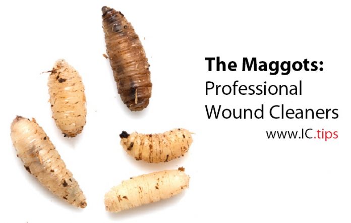
Maggot therapy is a biotherapy in which live, disinfected maggots (fly larvae) are placed in a non-healing wound to eat the necrotic tissue and disinfect the wound. Maggots have been used as a wound therapy since the beginning of civilization (1). Throughout the centuries, the benefits of maggots in wound healing have been repeatedly reported by surgeons in the army (2-6). During World War 1, William Baer, an orthopedic surgeon, noted the benefits of using maggots in compound fractures. He started maggot therapy at Hopkins hospital in Baltimore after a visit at the Boston’s Children Hospital (6).
The use of maggots declined with the advent of antibiotics. However, in the face of rising resistance to antibiotics, maggot wound therapy saw a revival when the FDA approved maggot therapy as a prescription only medical device for the debridement of necrotic tissue. They were classified as a device and not a drug, because the physical activity of the maggots was believed to be necessary for debridement (7). Maggots are used in the treatment of non-healing necrotic skin and soft tissue wounds, pressure ulcers, venous stasis ulcers and non-healing traumatic or post-surgical wounds (8-11). Research into maggot therapy has increased since FDA approval especially in Europe, leading to two published PhD theses from the Netherlands (12,13). In this review, we discuss the usage and benefits of maggot therapy.
four main actions of maggots on wounds
There are four main actions of maggots on wounds; debridement, disinfection, stimulation of healing, and biofilm inhibition and eradication (8-11). Most larvae are derived from the green bottle fly or bow fly, Lucilia (Phaenicia) sericata. Approximately 10 maggots are required per square centimetre of the wound surface. In maggot therapy these are sterilized larvae delivered in “biobags” or in bottles (14,15). The larvae are ordered one day before the start of treatment and are fed with physiological saline. The biobags are square and are used for the smaller wounds. They are placed at the wound and attached with a closing wound dressing. For bigger wounds the larvae are placed directly onto the wound surface. A median treatment of 3-4 days will cost € 150-160, with a price of € 160/bottle or pack of biobags (Monarch Labs LLC,Irvine, CA, USA). At the start of the treatment, the larvae are 1-2 mm in size and after 3-4 days, they grow to 10-20 mm in length. Wound fluids and liquefied necrotic tissue can enter the biobag and support the growth of the maggots. The maggots feed through a process known as extracorporal digestion. This is a process where maggots excrete digestive enzymes and feed off the liquefied material (also known as alimentary secretion and excretions, ASE). The physical movement of the maggots contribute significantly to the debridement of the wound (9,10.14).
The ASE proteolytic enzymes include a wide array of matrix-metalloproteinases (MMPs). They include trypsin-like and chymotrypsin-like serine proteases, an aspartyl protease, and an exopeptidase-like MMP (16-19). MMPs play critical roles in all phases of wound healing including hemostasis, thrombosis, inflammatory cell activation, collagen degradation, fibroblast and keratinocyte migration, and tissue remodelling. Disturbances in wound healing can occur when one group of proteases is deficient, or out of balance with another (16-19). Some proteases excreted by the maggots are resistant to wound proteases (20).
Evidence of Efficacy
Numerous studies about the efficacy of maggots have been published (8-11,21-23). In a 2002 study, Sherman et al studied maggot therapy versus control debridement therapy for pressure ulcers. The average surface area of necrotic tissue for wounds treated with maggot therapy (n=42) decreased faster and greater than that observed with conventional therapy (n=49). Of the 31 measurable conventional treated wounds necrotic tissue decreased 0.2 cm2 per week and total wound area increased 1.2 cm2 per week. During maggot therapy, necrotic tissue decreased 0.8 cm2 per week and total wound surface decreased 1.2 cm2 per week (24). Similarly, it was demonstrated that maggot debridement therapy for diabetic foot ulcers decreased the average surface area of necrotic tissue for wounds faster and greater compared to conventional therapy (n=14) (25). In a 2004 study, Sherman and Shimoda reported decreased rates of postoperative wound infections in patients receiving pre-surgical maggot debridement (n=10). Ten wounds were debrided with maggots for (1-17 days) prior to surgical closure. Postoperative wound infection was not observed. Six of the 19 wounds not treated with maggots developed postoperative wound infections (26). Additionally, between 2002 and 2005 several case reports were published reporting the benefits of maggot debridement therapy (MDT) in amputation sparing surgery and after breast-conserving surgery (27,28).
In 2009, the first randomized controlled trial of larval therapy in leg ulcers was published. The VenUS-2 trial was an open randomized trial with 267 patients with at least one venous or mixed venous-arterial ulcer and with at least 25% coverage of slough or necrotic tissue (8). It was a 3-armed trial comparing loose larvae, bag larvae and hydrogel. The primary outcome was time to healing of the largest eligible ulcer. Maggot therapy scored only better in the time to debridement, whereas for the secondary outcomes (including time to debridement, bacterial load, presence of methicillin-resistant Staphylococcus aureus (MRSA), quality of life, adverse effects, and ulcer related pain), no improvement was seen when maggots were utilized (8).
In an open letter to the editor of the BMJ, Sherman criticized the study design stating that the true significance of this study may be in demonstrating that the time saved by larval debridement may be lost if we do not continue to address the quality of the wound, and its response to treatment throughout the entire healing process. The fact that subjects in all three study arms failed to heal as quickly as expected, supported the contention, that the study design was not consistent with good clinical practice. In the opinion of Sherman, eight months to heal a 12 cm2 ulcer must not be accepted as the standard of care, (29).
In 2012, a randomized multicenter trial (n=119) compared two groups of patients receiving MDT or the conventional treatment for wound debridement during a two-week hospital stay. Percentages of slough were significantly lower in the MDT group at day 8, but not at day 15. The authors concluded that because there is no benefit in treatment after 1 week, another type of wound dressing should be used (30). In 2014, Mudge et al also noted faster debridement in favour of larval therapy over hydrogel therapy in an randomized, controlled trial of patients with leg ulcers (n=88) (31).
A Cochrane meta-analysis on the efficacy of MDT was conducted in 2015, which identified 10 studies with 715 participants. The overall quality of evidence was low, because the studies were small and most were short duration. There were mixed results between the studies in terms of the amount of slough in the wound bed at the start of the trial, the duration of the treatments, and the methods used to see how well the debridement worked (32). There is no strong evidence whether debridement itself or any particular form of debridement is beneficial in the treatment of venous ulcers (33,34).
Disinfection of the Wound
In insects, the most widespread antimicrobial peptides (AMPs) are defensins. Some medicinally important compounds, including lucifensin 1 and lucifensin 2 can potentially be used as synthetic AMP analogues in wound dressings (37). Maggots have been shown to have anti-bacterial properties. Huberman et al showed that the extracts and haemolymph of Luvilia sericata had bactericidal activity against Gram-positive and Gram-negative bacteria, including Pseudomonas aeruginosa, Klebsiella pneumonia and methicillin-resistant Staphylococcus aureus isolated from wounds (35). This bactericidal activity was attributed to a variety of AMPs released by the maggot larvae. Poppel et al. identified a whole spectrum of AMPs consisting through RNA sequencing, including lucifensin and lucimycin, attacins, cecropins, diptericins, proline-rich peptides and sarcotoxins. They identified 47 genes encoding putative AMPs and detected the presence 23 AMP analogues (36). Some analogues had activities against various pathogens, including Pseudomonas aeruginosa, Proteus vulgaris and Enterococcus faecalis. In some instances, synergistic effects were found against Escherichia coli and Micrococcus luteus, when selected AMPs combinations were used (36).
Stimulation of Wound Healing
Wound healing is a very complicated process in which a major cytoskeletal component, vimentin, acts as a signal integrator during wound healing; operating in both signalling delivery and response (38). It was believed for some years that the physical properties of the maggots were the most important for wound healing, because loose maggots healed wounds faster than maggots applied while in a bag/container. Current evidence suggests that the secretions components of the maggots are the most important (39,40). Reports from in vitro studies suggest that MDT could be replaced with application of the secreted against from maggots alone (39,40).
Complement activation(CA) is needed to initiate tissue repair. However, inappropriate activation of complement, as observed in chronic wounds, can cause cell death and enhance inflammation. In this way inappropriate CA contributes to further injury and impaired wound healing. Therefore, attenuation of CA by specific inhibitors may assist to facilitate wound healing. Currently the effects of several complement inhibitors, such as the C3 inhibitor compastin, and several C1 and C5 inhibitors are under investigation in wound healing (41,42). Cazander et al. showed maggot secretions reduce CA in healthy and post-surgical immune activated sera by up to up to 99,9% via all pathways. Maggot excretions do not specifically initiate or inhibit CA, but breakdown complement proteins C3 and C4 to attenuate CA (41,42).
Conclusion
Maggot wound debridement therapy has made a revival in the last two decades. However, the quality of research has been insufficient to establish its efficacy. In a Cochrane meta-analysis, the overall evidence for its use was considered low (32). Nevertheless, a clear and consistent pattern in all the studies suggest that the maggots increased the speed of wound debridement in the first week of treatment compared to hydrogel therapy (10,24,28). In addition, maggots have been shown to produce anti-microbial peptides, which have broad-spectrum bactericidal activity. These AMPs can be synthesized. They are able to reduce the complement system activity (41-42). The application of synthetic AMP analogues in wound dressings and adhesive bandages will potentially solve many issues in wound care (43).
References
- Whitaker I.S., Twine C., Whitaker M.J., et al. Larval therapy from antiquity to the present day. Mechanisms of action,clinical applications and future potential. Postgrad Med J. 2007;83;(980);409-13.
- Sherman R.A., Hall M.J., Thomas S. Medicinal Maggots: An Ancient Remedy for Some Contemporary Affictions. Ann Rev Entomol.2000;45;55-81.
- Donolly J. Wound healing: from poultices to maggots ( a short synopsis of wound healing throughout the ages) Ulster Med.J..1998;67;Suppl.1;47-51.
- Baer W.S. The treatment of chronic osteomyelitis with the maggot(larva of the bow fly) J Bone Joint Surg. 1931;(3);438-75.
- Heitkamp R.A., Peck G.W., Kirkup B.C. Maggot Therapy in Modern Army Medicine: Perceptions and Prevalence. Military Medicine 2013;177;(11);1411-16.
- Manting M.M., Calhoen M.D. Biographical Sketch: William S. Baer (1872-1931) Clin Orthop Rel Res. 2011;Apr;469(4);917-19.
- US Food and Drug Administration. Medical Maggots 2004;K 033391; Accessed Oct 10, 2017 https://www.accessdata.fda.gov/cdrh_docs/pdf7/K072438.pdf
- Dunville J.C., Bland M.J., Cullen N., et al. Larval therapy for leg ulcers (VenUS-2):randomized, controlled trial. BMJ 2009;338b773.
- Andersen A.S., Sandvang D., Schnorr K.M. A novel approach to the antimicrobial activity of maggot therapy. J Antimicrob Chemother.2010;Aug;65;(8);1646-54.
- Nishijima A., Gosho M., Yoshida R., et al. Effective wound bed preparation using maggot debridement therapy for patients with critical limb ischaemia. J Wound Care 2017;26;8;483-89.
- Serra M.B., Barroso W.A., Neves da Silva N., et al. From Inflammation to Current and Alternative Therapies Involved in Wound Healing. Int J Inflamm. 2017;Art.ID; 3406215.
- Steenvoorde P. Maggot Debridement Therapy in Surgery. PhD. Thesis Leiden University, 2008, Jan.9th.
- Cazander G. Wound Healing, Larval Therapy. PhD. Thesis Free University, Amsterdam,2010.
- Fleischmann W., Grassberger M., Sherman R.A. Therapy: A Handbook of Maggot –Assisted Wound Healing. Thieme Verlag, Stuttgart,2004;1-93.
- Grassberger M., Fleischman W. The Biobag: A New Device for the Application of Medicinal Maggots. Dermatol. 2002;204;306.
- Vistmes L.M., Lee R.,Kasander G.A. Proteolytic activity by bow fly larvae secretions in experimental burns. Surgery 1981;90(5);835-41.
- Telford G., Brown A.P., Seabra R.A., et al. Degradation of eschar from venous leg ulcer using a recombinant chymotrypsin from Lucilia sericata. Br.J.Derm. 2010;163(3);523-31.
- Gill S.E., Parks W.C. Metalloproteinases and their inhibitors: regulation of wound healing. Int J Biochem Coll Biol. 2008;40(6-7);1334-47.
- Pritchar D.I., Telford G., Diab M., et al. Expression of cGMP compatible Lucilia sericata insect serine protease debridement enzyme. Biotechnol Prog. 2012:28;(2);567-72.
- Telford G., Brown A.P., Kind A. Maggot Chymotrypsin 1 from Lucilia sericata is resistant to endogenous wound protease inhibitors. Br J Derm. 2011;164;192-96.
- Turner J.T. Debriding Chronic Wounds with Larval Therapy: The New Answer for Biosurgery in the Outpatient Wound Clinic? Wound Clinic 2017; Jul;11;7.
- Sun X., Jiang K., Chen J. A systematic review of maggot debridement therapy for chronically infected wounds and ulcers. Int J Inf Dis. 2014;25;32-37.
- Nigam Y. Advances in Myasis Treatment. Health Care Curr Rev. 2016;4;161
- Sherman R.A. Maggot therapy versus conservative debridement therapy for the treatment of pressure ulcers. Wound Repair Regen. 2002;10;208-14.
- Sherman R.A. Maggot therapy for treating diabetic foot ulcers unresponsive to conventional therapy. Diabetes Care 2003;26(2);446-51.
- Sherman R.A., Shimoda K.J. Presurgical Maggot Debridement of Soft Tissue Wounds is Associated with Decreased Postoperative Infections. Clin.Inf.Dis.2004:38(7);1067-70.
- Jukema G.N., Menon A.G., Steenvoorde P., et al. Amputation sparing surgery by nature: maggots revisited. Clin Inf Dis. 2002;35(12);1566-71.
- Steenvoorde P., Oskam J. Use of larval therapy to combat infection after breast-conserving therapy. J Wound Care 2005;14(5);212-13.
- Sherman R.A., et al. Maggot Therapy: Apparently a Good Treatment Despite Poor Study. BMJ 2009;338.
- Opletalova K., Blaizot X., Mourgeon B., et al. Maggot Therapy for Wound Debridement: A Randomized Trial. Arch Dermatol 2012;148(4);432-38.
- Mudge E.M., Price P., Neal W, et al. A randomized controlled trial of larvae therapy for the debridement of leg ulcers; Results of a multicenter randomized, contolled, open, observer blind, parallel Group study. Wound Repair Regen. 2014;22;43-51.
- Cowman G.C., Kolback D.N. Debridement for venous leg ulcers. Cochrane Library: Wound Group.2015; Issue 9; Art.No.CD008599.
- Barnes S., Spencer M., Graham D. Surgical wound irrigation: a call for evidence-based standardization of practice. Am J Infect Control. 2014; May;42(5);525-29.
- The Flow Investigators. A Trial of Wound Irrigation in the Initial Management of Open Wounds. New Engl J Med. 2015;373;2629-41.
- Huberman L., Gollop N., Mumcuoglu K.Y.,et al. Antimicrobial properties of whole body-extracts and haemolymph of Lucilia sericata maggots. J Wound Care 2007; Mar;16(3);123-27.
- Poppel A.K., Vogel H. Wiesner J., et al. Antimicrobial Peptides Expressed in Medicinal Maggots of the Bow Fly, Lucilia sericata Show Combinatorial Activity Against Bacteria. Antimicrob.Agents Chemother. 2015; May;59(5); 2508-14.
- Celrovsky V., Bem R. Lucifensins: The Insect Defensins of Biomedical Importance; The Story Behind Maggots Therapy. Pharmaceuticals 2014;7(3);251 -64.
- Cheng F., Shen Y., Mohanasundaram P., et al. Vimentin coordinates fibroblast proliferation and keratinocyte differentiation in wound healing via TGF-beta-Slug signalling. Proc Natl Acad Sci. USA 2016;113(30); E4320- 27.
- Smith A.G., Powis R.A., Pritchard D.J., et al. Greenbottle (Lucilia sericata) larval secretions delivered from a prototype hydrogel wound. Biotechnol Prog. 2006;22(6); 1690-96.
- Cazander G., GottrupF., Jukema G.N. Maggot therapy for wound healing: clinical relevance, mechanisms of action and future prospects. J Wound Techn. 2009;5(18);2321.
- Cazander G., Jukema G.N., Nibbering P.H. Complement Activation and Inhibition in Wound Healing. Clin Develop. Immunol. 2012. 534291;1-18.
- Cazander G., Schreurs M.W., Renwarin L., et al. Maggot excretions affect the human complement system. Wound Repair Regen. 2012;20;879-86.
- Grönberg A., Maluapua M., Stähle M., et al. Treatment with LL-37 is safe and effective in enhancing healing of hard-to-heal venous leg ulcers: a randomized placebo-controlled trial. Wound Repair Regen 2014; 22;613-21.


